Foot Muscles Mri - Ankle and Foot | Radiology Key

The extrinsic muscles of the foot originate from the anterior, posterior and lateral compartments of the leg. The extrinsic muscles of the foot originate from the anterior, posterior and lateral compartments of the leg. Human anatomy for muscle, reproductive, and skeleton. Indications for foot mri scan. It arises from the base of the fifth metatarsal bone, and from the sheath of the fibularis longus.
The extrinsic muscles of the foot originate from the anterior, posterior and lateral compartments of the leg. Upper and lower lines mark. Muscles of the foot are located on its rear and on the sole. Learn vocabulary, terms and more with flashcards, games and other study tools. The muscles acting on the foot can be divided into two distinct groups;
The muscles acting on the foot can be divided into two distinct groups;
The muscles acting on the foot span from above the knee to various points on the foot skeleton. Learn vocabulary, terms and more with flashcards, games and other study tools. Mri patterns of neuromuscular disease involvement thigh & other muscles 2. ► shoulder ► elbow ► wrist ► finger ► thumb. Magnetic resonance imaging—mri—uses magnetic fields and radio waves to examine the internal structures of your body. Mri with hardware in foot? Subscribe to foot & ankle problems. Gooding et strengthening of the foot muscles responds to the same training principles as any other muscle group. ► hip ► pelvis ► thigh ► knee ► lower extremity/shin ► ankle ► foot. Foot positioned for axial images of the ankles;
These muscles begin and attach within the skeleton of the foot, have complex anatomical and topographical and functional relationships with. Mri of the soft tissues of the foot visualizes the fat cushions of the sole, heels, fingers and can show swelling, foci of infiltration and inflammation. Bone contusions, osteonecrosis, marrow oedema syndromes, and stress > fractures) > synovial based disorders ( e.g.
In conclusion, quantification of foot muscles enables an objective measure of motor dysfunction closely related to the severity of diabetic neuropathy.
Learn vocabulary, terms and more with flashcards, games and other study tools. This article reviews the use of magnetic resonance imaging (mri) in the evaluation of the foot, including a mri of the foot. Related posts of foot muscle anatomy mri. Indications for foot mri scan. Bone contusions, osteonecrosis, marrow oedema syndromes, and stress > fractures) > synovial based disorders ( e.g. Abdm, abductor digiti minimi muscle; The muscles with proximal attachments at points outside the foot are referred to as extrinsic. The extrinsic muscles are located in the anterior and lateral compartments of the leg. Mri with hardware in foot? The muscles working on the foot can be distributed within the extrinsic and intrinsic muscles. Posted by radiologyer at 8:12 am. In addition, an image of all the muscles of the back and.
.magnetic resonance imaging (mri) or ultrasound imaging (usi) (soysa et al., 2012; Muscles of the foot muscle origin insertion nerve supply extensor digitorum brevis distal part of the lateral and superior surfaces of the calcaneus and the apex of the inferior extensor. Related posts of foot muscle anatomy mri. Learn vocabulary, terms and more with flashcards, games and other study tools. Indications for foot mri scan. Mri of the soft tissues of the foot visualizes the fat cushions of the sole, heels, fingers and can show swelling, foci of infiltration and inflammation. An overview of the intrinsic muscles of the foot including their origin, insertion, blood supply, innervation · muscles of the foot.

► shoulder ► elbow ► wrist ► finger ► thumb.
Bone contusions, osteonecrosis, marrow oedema syndromes, and stress > fractures) > synovial based disorders ( e.g. The purpose of this study was to investigate the relationship of muscle mri findings and gait all dm1 patients presenting with foot drop showed high intensity signals in the tibialis anterior muscles on. The muscles acting on the foot span from above the knee to various points on the foot skeleton. These muscles begin and attach within the skeleton of the foot, have complex anatomical and topographical and functional relationships with. By muhammad ali, mb bs; ► shoulder ► elbow ► wrist ► finger ► thumb. Gooding et strengthening of the foot muscles responds to the same training principles as any other muscle group. Mri with hardware in foot? Indications for foot mri scan. The second part is on the plantar group of muscles. Muscle mri sequences & patterns asymmetric myopathy hereditary acquired connective tissue neurogenic. .magnetic resonance imaging (mri) or ultrasound imaging (usi) (soysa et al., 2012; The deformity of the foot with abnormal pressure distribution on the plantar surface coupled with reduced or loss of sensation, makes the foot.
 was performed on a normal subject;")
Hi, i had surgery on my shoulder about 8 years ago and have two metal anchors in my shoulder.
 was performed on a normal subject;")
The flexor digiti minimi brevis (flexor brevis minimi digiti, flexor digiti quinti brevis) lies under the metatarsal bone on the little toe, and resembles one of the interossei.

Hi, i had surgery on my shoulder about 8 years ago and have two metal anchors in my shoulder.

By muhammad ali, mb bs;

Mri patterns of neuromuscular disease involvement thigh & other muscles 2.

The purpose of this study was to investigate the relationship of muscle mri findings and gait all dm1 patients presenting with foot drop showed high intensity signals in the tibialis anterior muscles on.

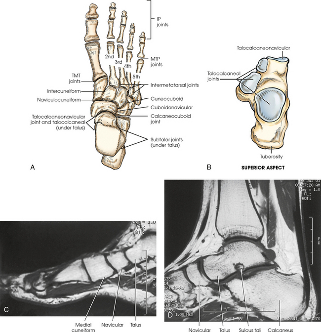
A magnetic resonance imaging (mri) was performed on a normal subject;

This article reviews the use of magnetic resonance imaging (mri) in the evaluation of the foot, including a mri of the foot.

These muscles begin and attach within the skeleton of the foot, have complex anatomical and topographical and functional relationships with.

Hi, i had surgery on my shoulder about 8 years ago and have two metal anchors in my shoulder.

Bone contusions, osteonecrosis, marrow oedema syndromes, and stress > fractures) > synovial based disorders ( e.g.

In addition, an image of all the muscles of the back and.
 lies under the metatarsal bone on the little toe, and resembles one of the interossei.")
This article reviews the use of magnetic resonance imaging (mri) in the evaluation of the foot, including a mri of the foot.

Mri with hardware in foot?

Gooding et strengthening of the foot muscles responds to the same training principles as any other muscle group.

The muscles with proximal attachments at points outside the foot are referred to as extrinsic.

Indications for foot mri scan.

If you'd like to support us and get something great in return.

Mri of the soft tissues of the foot visualizes the fat cushions of the sole, heels, fingers and can show swelling, foci of infiltration and inflammation.

By muhammad ali, mb bs;

If you'd like to support us and get something great in return.

Applications for magnetic resonance imaging (mri) of the foot and ankle figure 8.4 image planes for foot and ankle mri.

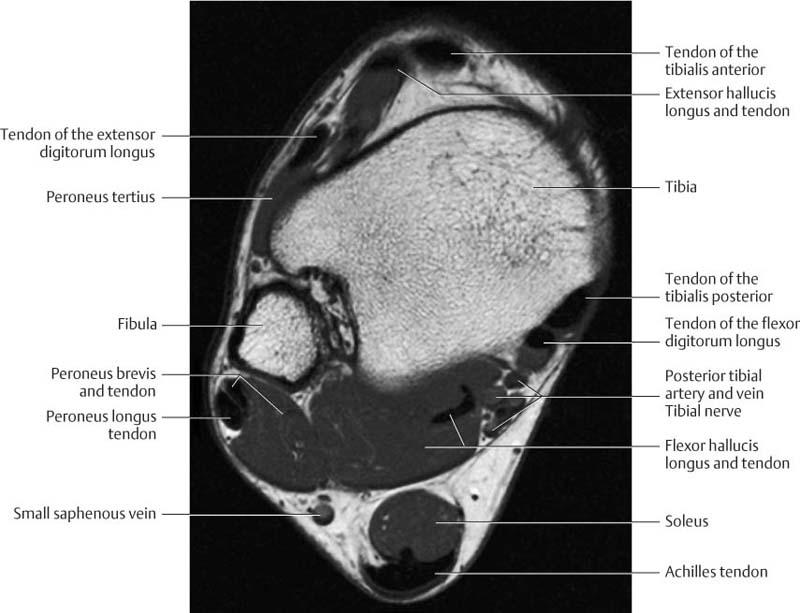
Foot positioned for axial images of the ankles;

Posted by radiologyer at 8:12 am.

Related posts of foot muscle anatomy mri.

► shoulder ► elbow ► wrist ► finger ► thumb.

Lateral and medial processes of calcaneal tuberosity.

Abdm, abductor digiti minimi muscle;
 was performed on a normal subject;")
In addition, an image of all the muscles of the back and.

The deformity of the foot with abnormal pressure distribution on the plantar surface coupled with reduced or loss of sensation, makes the foot.

Foot positioned for axial images of the ankles;
The deformity of the foot with abnormal pressure distribution on the plantar surface coupled with reduced or loss of sensation, makes the foot.

Mri with hardware in foot?

Start studying mri procedures foot/ankle review.

The flexor digiti minimi brevis (flexor brevis minimi digiti, flexor digiti quinti brevis) lies under the metatarsal bone on the little toe, and resembles one of the interossei.

Posting Komentar untuk "Foot Muscles Mri - Ankle and Foot | Radiology Key"